
Neuron and Axon
http://prettynerdygirls.blogspot.com/2011/04/neurons-and-axons.html
Cranial Nerves (Fixed!!)
http://prettynerdygirls.blogspot.com/2011/04/cranial-nerves.html
Osteon models and Tarsal Bones
http://prettynerdygirls.blogspot.com/2011/03/human-anatomy-details.html
Carpals, Bones, Skin
http://prettynerdygirls.blogspot.com/2011/03/no-answers-time-for-bed.html
Above: Figure 1
Below: Figure 2
Below: Figure 2
Above: Figure 3
Below: Figure 4
Above: Figure 5
Below: Figure 6
Below: Figure 6
Above: Figure 7
Below: Figure 8
Below: Figure 8
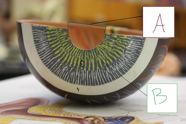
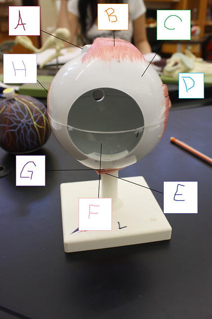
Figure 1:
A: Superior Oblique
B: Superior Rectus
C: Sclera
D: Lateral Rectus
E: Inferior Oblique
F: Corneum
G: Inferior Rectus
H: Medial Rectus
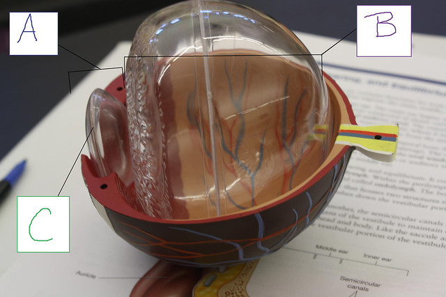
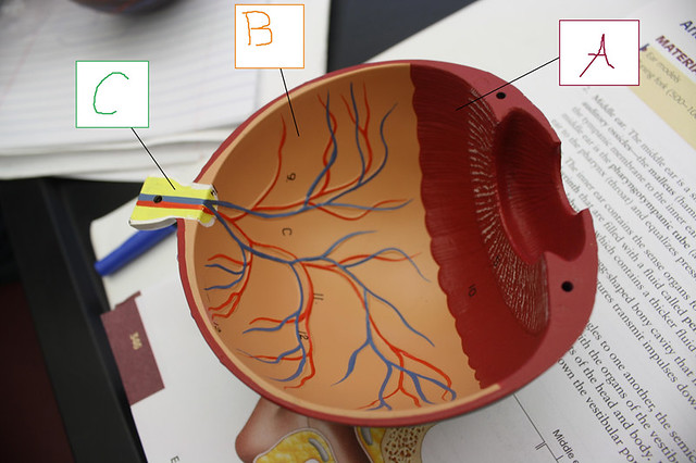
Figure 2:
A: Anterior Chamber
B: Posterior Chamber
C: Lens
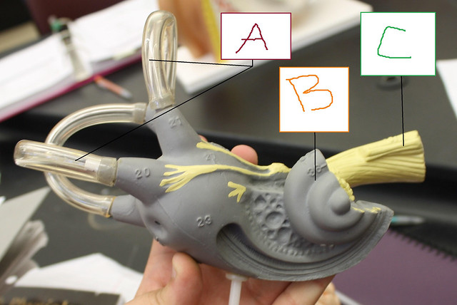
Figure 3:
A: Semicircular canals
B: Cochlea
C: Vestibulocochlear nerves
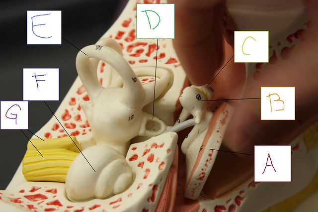
Figure 4:
A: Tympanic Membrane
B: Malleus
C: Incus
D: Stapes
E: Semicircular canals
F: Cochlea
G: Vestibulocochlear nerves
Figure 5:
A: Ciliary Body
B: Retina
C: Optic Nerve
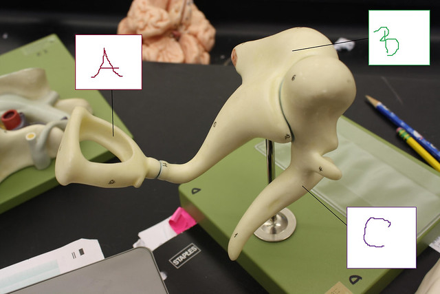
Figure 6:
A: Stapes
B: Incus
C: Malleus
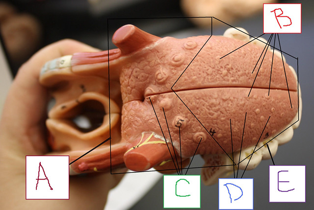
Figure 7:
A: Innervated by Glossopharyngeal IX
B: Fungiform Papillae
C: Circumvallete Papillae
D: Filiform Papillae
E: Innervated by Facial VII
Figure 8:
A: Pupil
B: Iris
No comments:
Post a Comment